Dr. Arlo Miller first became interested in dermatology while working in a a melanoma research lab at the Dana Farber Cancer Institute in Boston. His PhD dissertation at Harvard University focused on gene expression in melanomas. Treatment of melanoma and understanding melanoma biology remains one his primary interests.
Important Facts about Melanoma
- Although less common than other skin cancers, it causes far more deaths.
- There are about 120,000 new cases each year in the United States.
- About 70,000 of these cases are invasive melanoma.
- Early melanomas that are not invasive are curable with surgery.
- There are about 9,000 deaths from melanoma per year in the US from invasive melanomas.
Basic Biology of Melanoma
Melanoma refers to a type of skin cancer that comes from the cells in our body that make pigment. These specialized cells, called melanocytes, make up just a tiny fraction of the cells in our skin. Depending on a variety of genetic factors, they are programmed to make a certain type and quantity of pigment that determines our skin and hair color. Melanocytes will also churn out more pigment leading to a tan when their DNA is damaged or mutated by ultraviolet radiation from the sun or tanning beds.
Risk Factors for Melanoma
There are a variety of factors that affect the risk of developing melanoma. The most important factors are the amount of ultraviolet light exposure from the sun or tanning beds you have had, your ethnicity, having atypical moles, and having a personal or family history of melanoma. These factors can be divided into risks that you can control and ones that you cannot.
Controllable Risk Factors for Melanoma
Tanning Beds, Sun Tanning, and Melanoma Risk
Large studies have shown that tanning 10 or more times in a single year prior to the age of 30 has a 7.7x higher risk of melanoma. Even having gone to a tanning bed just once before the age of 35 leads to a 1.22x higher risk of melanoma. The tanning bed industry has tried to claim that tanning beds are safe, and has even claimed that it can protect against cancer. Meanwhile, the World Health Organization has classified tanning beds as a carcinogen delivery device because of the abundant data that ultraviolet radiation is the primary environmental factor causing skin cancer. Studies have shown that there is absolutely no truth to the claim that some types of tanning beds are safe and a recent congressional report that investigated 300 tanning parlors found that they routinely provide false and misleading information, particularly to children. More about tanning can be found here.
Sunburns, Intermittent Sun Exposure, and Why Washington State Has a High Rate of Melanoma
Your history of sunburns affects your risk of melanoma. Having one blistering sunburn under the age of 20 makes you 2x more likely to develop melanoma in your lifetime. Having three or more blistering sunburns under the age of 20 makes you 5x more likely.
However, its also been found that intense intermittent exposure, sort of like summers in Washington also raises your risk. This is probably the reason why Washington, despite being a northern state with less sun overall than someplace like Arizona, is in the CDC’s highest category of melanoma risk at 21.6-28-8 cases per 100,000 people. It is why its important to be diligent about sun safety around here.
Uncontrollable Risk Factors for Melanoma
Risk of melanoma based on skin color
Skin color has a major impact on the overall risk of melanoma. Since you can’t choose your parents, it is a melanoma risk factor that you cannot control. Based on data from 2009, you can see that those with darker skin color have much less risk:
- White 1:50
- Hispanic 1:250
- Native American 1:350
- Asian 1:800
- African American 1:1100
The same genes that control skin color are also related to hair and eye color and freckling. People with red or blond hair, blue or green eyes, or a tendency to freckle have a 2-3 times higher risk of melanoma.
It is widely thought that during human evolution, having heavily pigmented skin provided a tremendous advantage in equatorial areas by protecting people from skin cancer. However, in more temperate climates, heavy pigmentation prevents vitamin D synthesis and became a disadvantage. Today we still see the effects of this. African Americans living in the northern United States often have vitamin D deficiencies while Caucasians living in southern states have high rates of skin cancer.
Risk of melanoma based on how many moles you have and what they look like
The moles that we all have (pretty much EVERYONE has at least one somewhere) can be divided into two categories based on their dermoscopic appearance: normal (typical) or abnormal (atypical).
Having normal moles has very little impact on melanoma risk. If you have more than 50 normal moles, however, there is an increased risk of about 2-4x.
However, having atypical moles does bring a lot more risk. Just one atypical mole brings a risk of 2x. Having ten or more atypical moles brings a 12-14x risk. Having many atypical moles and a family member who has had a melanoma brings a lifetime melanoma risk of 100%.
This is why it is probably worth it for people worried about their moles to have a complete skin exam. Using dermoscopy, we can determine whether your moles are normal or atypical. Combined with other factors like your personal and family history, a recommendation can be made as to whether you could benefit from ongoing surveillance examinations.
Risk of melanoma based on personal or family history
If you have previously had a melanoma, your melanoma risk goes up by 9-28x. Specifically, this refers to getting a new melanoma, not having the previous one come back.
If you have had one of the non-melanoma types of skin cancer (basal cell or squamous cell carcinoma), your risk of melanoma is 3-5x higher.
Other unusual risk factors for melanoma
A variety of the other factors can increase melanoma risk, but don’t come up very often:
- On an immunosuppressant medicine 4-8x
- Having lupus or xeroderma pigmentosum Very High
What does melanoma look like?
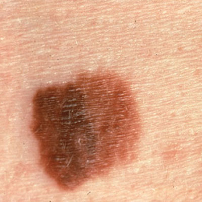
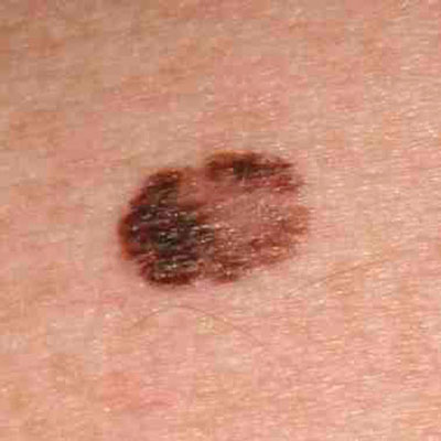
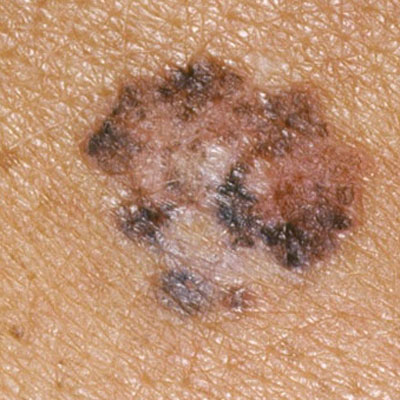
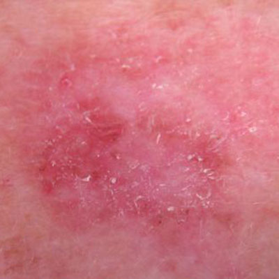
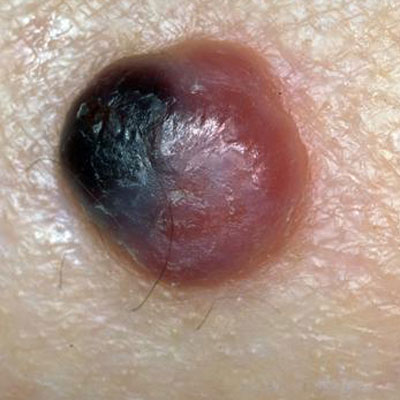
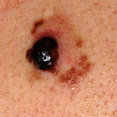
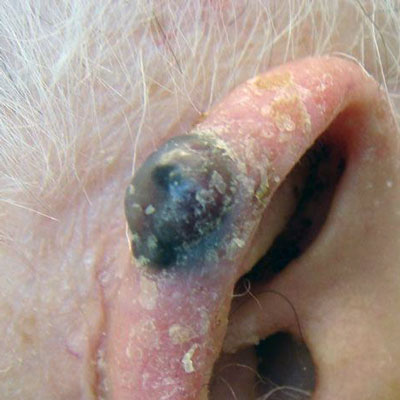
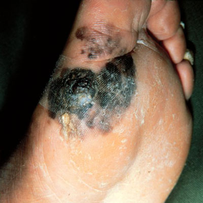
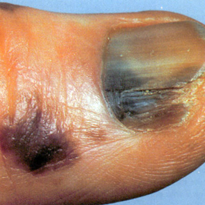
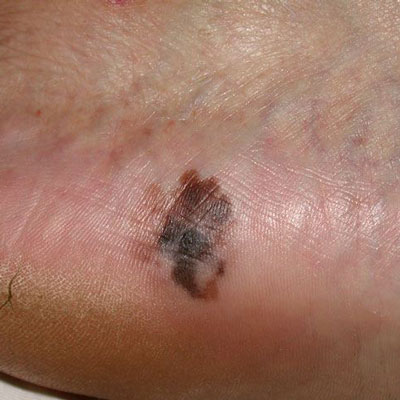
Melanomas often resemble moles, but typically are darker in color, have several shades of brown, have irregular edges, and are larger than an eraser. Another way to think of them is that normal or benign moles often look well put together, or orderly in some way, while melanomas often look like they were made by a slob. However, there are many exceptions to this general guide and there are melanomas that no longer make any brown pigment called a melanotic melanomas and are pink in color. These are much more difficult to detect. There are also melanomas that are pink and light brown in color. This tends to happen more often in redheads and is probably because they just don’t make dark brown pigment easily at all.


